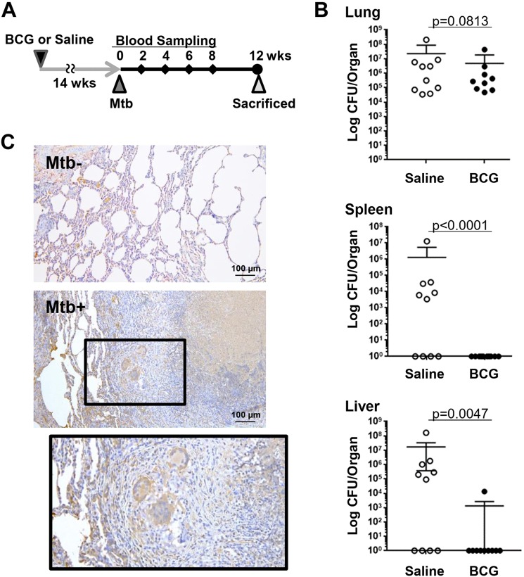
Figure 1.
A non-human primate model of tuberculosis. (A) Cynomolgus macaques were pre-treated with saline (n = 10) or BCG (n = 10) and challenged by Mtb. After the collection of blood samples at 0, 2, 4, 6, 8 weeks after Mtb challenge, monkeys were sacrificed for analysis at 12 weeks. (B) Bacterial burden of organs in macaques with or without BCG pretreatment at 12 weeks after Mtb challenge. Indicated organs were harvested 12 weeks after Mtb infection. Live Mtb counts (CFU) in organs were determined by the mycobacterial culture of organ homogenates. Bars represent mean ± SEM. *p < 0.05, by Mann-Whitney’s U test. (C) Immunohistochemical analysis of LRG. Paraffin sections of lung tissues obtained from Mtb-unchallenged (Mtb−) and Mtb-challenged (Mtb+) macaques were used for immunohistochemical analysis to visualize LRG protein expression in tuberculosis lesions.

