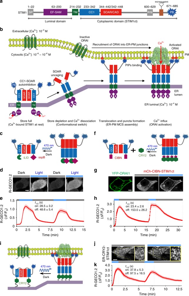
Fig. 1. Optogenetically engineered STIM1 permits light-switchable activation of ORAI Ca2+ channels.
Photostimulation was applied at 470 nm (4.0 mW/cm2). Data were shown as mean ± sem. Scale bar, 5 µm. a Domain architecture of the human STIM1. SP, signal peptide; EF-SAM, EF-hand and sterile alpha-motif; TM, transmembrane domain; CC1, coiled-coil domain 1; SOAR, STIM-Orai activating region; P/S, proline/serine-rich region; TRIP, the S/TxIP microtubule-binding motif; PB, polybasic tail. b Schematic of STIM1–ORAI1 coupling at the ER–PM junction that mediates store-operated Ca2+ entry. c–e Use of the iLID-sspB optical dimerizer to trigger STIM1ct activation and Ca2+ influx through endogenous ORAI channels. c Schematic of the design. iLID or sspB was fused to the N-terminus of STIM1ct at residue 233. d Confocal images showing photoswitchable Ca2+ influx in HeLa cells co-transfected with a red Ca2+ sensor (R-GECO 1.2) and the iLID/sspB fused STIM1ct chimeras. Cells were exposed to two repeated dark-light cycles. e Quantitative analysis of Ca2+ signals in response to repeated photostimulation (n = 40 cells from three independent experiments). The half-lives (t1/2) of on and off kinetics were fitted with one phase exponential decay (“±” means 95% confidence interval). f–h Use of the CRY2-CIBN optical dimerizer to photo-activate STIM1ct and Ca2+ influx. f Schematic of the design. CRY2 was used to photo-crosslink CIBN-STIM1ct and trigger STIM1ct activation to induce Ca2+ entry. g Confocal images showing light-induced co-localization of mCherry (mCh)-tagged CIBN-STIM1ct with YFP-ORAI1 in HeLa cells. h Reversible Ca2+ responses monitored by R-GECO 1.2 (n = 30 cells). Blue bar, photostimulation at 470 nm with a power density of 4 mW/cm2. i–k ER-tethered CRY2-STIM1ct mimics STIM1 puncta formation at ER–PM junctions to evoke localized Ca2+ influx. i Schematic of the design. j Confocal images illustrating light-induced clustering of ER-resident CRY2-STIM1ct at the footprint of HeLa cells. Enlarged views of the boxed regions were shown on the right. k Cytosolic Ca2+ signals reported by R-GECO1.2 in HeLa cells subjected to two repeated dark-light cycles (n = 30).

