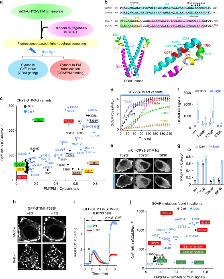
Fig. 5. An optogenetic platform for screening of STIM1 gain- or loss-of-function mutations.
Data were shown as mean ± sem. Scale bar, 5 µm. a Design of the high-throughput screening pipeline. The cytosol-to-PM translocation and Ca2+ influx (GCaMP6s as readout) were used as two readouts. b Sequence alignment of human SOAR1 and SOAR2 domains and the 3D structure of SOAR1 (PDB entry: 3TEQ). Key residues at the interdimer interface or involved in ORAI1-binding were indicated by dots and triangles, respectively. Selected key residues were highlighted in the 3D structure. c Quantification of Ca2+ responses (GCaMP6s) and PM translocation (mCherry signals) of selected CRY2-STIM1ct mutants before (dark dots) and after (blue dots) photostimulation. HeLa-GCaMP6s stable cells were co-transfected with each of the indicated mCh-CRY2-STIM1ct mutants and ORAI1-CFP. d Time courses showing the kinetics of light-induced Ca2+ influx for WT and the indicated mCh-CRT2-STIM1ct variants. n = 60 cells. e–g Representative confocal images (e) and quantification of intracellular Ca2+ signals, n = 60 cells. Box-whisker plots indicated the median, and the interquartile range with 5–95 percentile distribution. f, as well light-induced PM translocation, n = 8 cells (g), in HeLa cells expressing WT or the indicated mCh-CRY2-STIM1ct mutants. h Representative confocal images of HEK293 S1-KO cells expressing the GFP-tagged full-length STIM1-T393F mutant before and after TG-induced store depletion. i SOCE monitored by R-GECO1.2 in HEK293 S1-KO cells expressing GFP-STIM1 WT or the mutant T393F. n = 90 cells. j Summary of the degrees of Ca2+ influx and PM translocation of cancer-associated mutations found in the SOAR domains of STIM1. HeLa cells were transfected with the indicated mCh-CRT2-STIM1ct mutants. Gain-of-function (H395Y and R424W; red) and loss-of-function (L402R, R426L/C, R429C; green) mutations were both identified. n = 60 cells.

