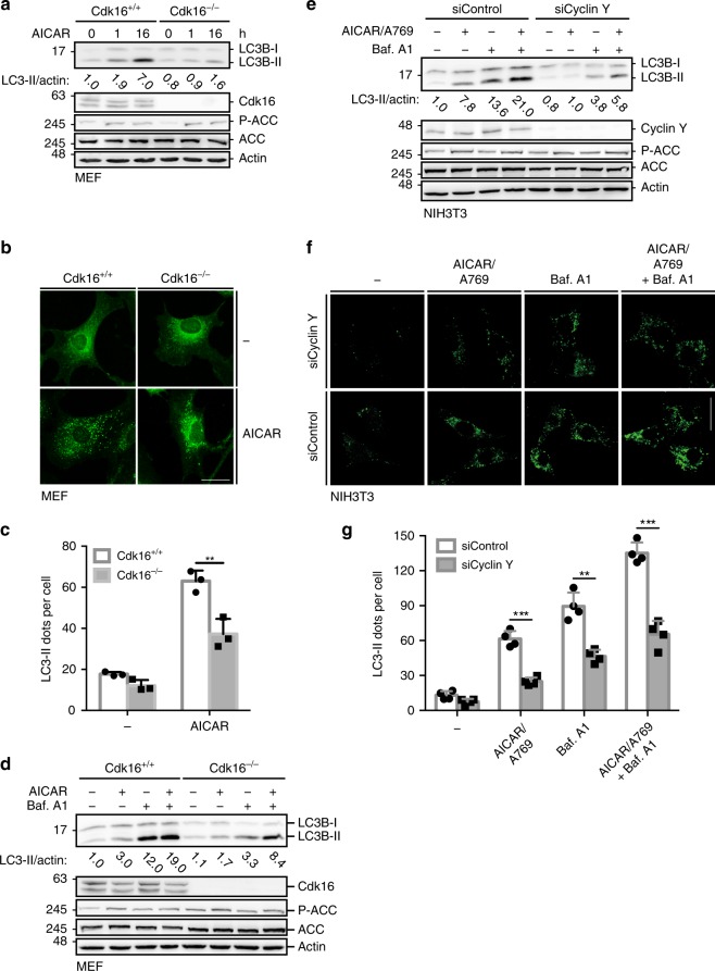
Fig. 6. AMPK-induced autophagy requires Cyclin Y/CDK16.
a Immortalized Cdk16+/+ and Cdk16−/− MEFs were treated with 1 mM AICAR for 1 or 16 h. Lysates were immunoblotted with the indicated antibodies (n = 3). b Representative confocal images of the Cdk16+/+ and Cdk16−/− MEFs treated as in panel a. Endogenous LC3 Staining with the 4E12 antibody monitored autophagy. Scale bar: 50 µm. c LC3 dots of experiments as shown in panel b were quantified. Statistical significance was measured via unpaired and two-tailed Student’s t-tests and is presented as follows: **p < 0.01. All error bars indicate SD. (n = 3; 100 cells counted for each replicate; Cdk16+/+ + AICAR vs. Cdk16−/− + AICAR: t = 4.964; df = 4). d Immortalized Cdk16+/+ and Cdk16−/− MEFs were treated with 1 mM AICAR for 1 h and/or with 200 nM Bafilomycin A1 (Baf. A1) for 6 h as indicated. For the combination, AICAR was added during the last hour of Baf. A1 treatment. Lysates were immunoblotted with the indicated antibodies (n = 1). e NIH3T3 cells were transfected with siRNA against Cyclin Y or control siRNA and stimulated with 0.5 mM AICAR/50 µM A769662 (A769) or with 200 nM Baf. A1 or a combination. Proteins were analyzed as indicated. (n = 3). f Representative confocal images of the NIH3T3 cells treated as in panel e. Endogenous LC3 was stained with the 4E12 antibody to depict autophagy. Scale bar: 50 µm. g LC3 dots as shown in panel f were quantified. Statistical significance was measured via unpaired and two-tailed Student’s t-tests and is presented as follows: **p < 0.01, ***p < 0.001. All error bars indicate SD. (n = 3; 100 cells counted for each replicate; AICAR/A769: t = 9.494, df = 6; Baf. A1: t = 6.549, df = 6; AICAR/A769 + Baf. A1: t = 9.484, df = 4). n biological independent replicate. SD standard deviation. Source data are provided as a Source Data file.

