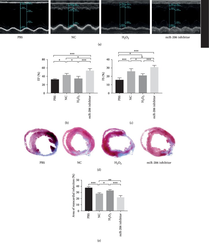
Figure 7.
Downregulation of miR-206 in MSCs effectively preserved cardiac function. (a) Representative images of rat heart function detected by echocardiogram in PBS, NC, H2O2, and miR-206 inhibitor groups at 28 days after MI (n = 5). The left ventricular ejection fraction (EF) (b) and fraction shorting (FS) (c) in the four groups were statistically analyzed (n = 5). (d) Representative images of Masson trichrome (MT) staining in PBS, NC, H2O2, and miR-206 inhibitor groups at 28 days after MI. (e) Percentage of fibrotic area calculated and analyzed by ImageJ software (n = 5). ∗P < 0.05, ∗∗P < 0.01, and ∗∗∗P < 0.001.

