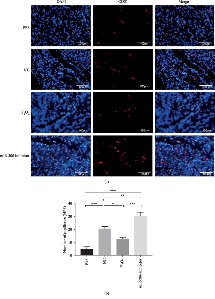
Figure 8.
CD31 staining of angiogenesis in rat hearts after MI. (a) Representative images of neovascular in rat heart infarction area in PBS, NC, H2O2, and miR-206 inhibitor groups detected by CD31 staining (n = 3). (b) CD31-positive vessels in the four groups were statistically analyzed (n = 3). ∗P < 0.05, ∗∗P < 0.01, and ∗∗∗P < 0.001.

