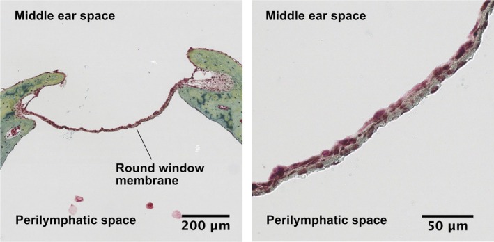
Figure 2.

Cross section of the guinea pig RWM, pentachrome stain. The RWM is suspended between bone, which is stained in green, shown on the left at ×8 magnification. The three layers of the RWM can be appreciated at ×40 magnification, shown on the right. The outer epithelial layer, in contact with the middle ear space, is composed of low cuboidal epithelial cells connected by tight junctions. The central connective tissue layer is stained beige due to its composition of collagen (yellow) and elastic fibers (black). The inner epithelial layer is composed of squamous epithelial cells with large extracellular spaces allowing contact between the connective tissue matrix and the perilymph. RWM, round window membrane. Printed with permission from Jeffrey W. Kysar, PhD and Anil K. Lalwani, MD
