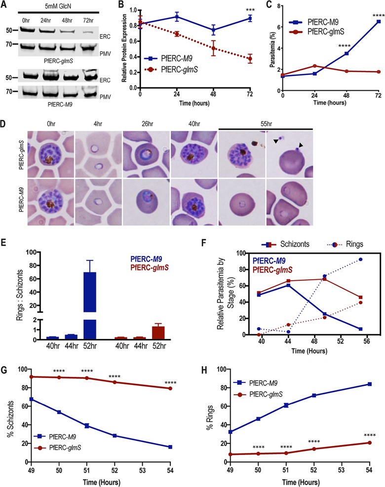
FIG 2.
PfERC mutants fail to transition from schizonts to rings. (A) Western blot of parasite lysates isolated from PfERC-glmS and PfERC-M9 parasites grown in the presence of 5 mM GlcN and probed with anti-HA antibodies and anti-plasmepsin V (PMV) antibodies. Results of one representative experiment of four are shown. (B) Quantification of changes in expression of PfERC in PfERC-glmS and PfERC-M9 parasites after addition of GlcN, as described for panel A. Data were normalized to the loading control (PMV) and are shown as means ± standard errors of the means (n = 4 biological replicates; ***, P < 0.001 [2-way analysis of variance {ANOVA}]). (C) Growth of asynchronous PfERC-glmS and PfERC-M9 parasites incubated with 5 mM GlcN, over 4 days, was observed using flow cytometry. Data are presented as means ± standard errors of the means (n = 3 technical replicates; ****, P < 0.0001 [2-way ANOVA]). One representative data set from 4 biological replicates is shown (Fig. S2A). (D) Representative Hema-3-stained blood smears of synchronous PfERC-glmS and PfERC-M9 parasites grown in the presence of GlcN (n = 2 biological replicates). (E) GlcN was added to synchronous PfERC-glmS and PfERC-M9 schizonts, and parasite stages were determined using flow cytometry. The ratio of rings to schizonts was calculated using the number of rings and schizonts observed at each time point. Data are presented as means ± standard errors of the means. (F) Hema-3-stained blood smears of synchronous PfERC-glmS and PfERC-M9 parasites grown in the presence of GlcN (shown in panel D) were manually counted. The amount of each life cycle stage (ring, trophozoite, and schizont) was determined as a percentage of the total number of parasites for each time point. (G and H) GlcN was added to synchronous PfERC-glmS and PfERC-M9 schizonts, and parasite stages were determined for schizonts (G) and rings (H) by the use of flow cytometry. At each time point, cells were fixed and stained with the DNA dye Hoescht 33342 to distinguish between ring stage parasites (1 N) and schizont stage parasites (16 to 32 N). Results of one representative experiment of three biological replicates are shown. Data are presented as means ± standard errors of the means (n = 3 technical replicates; ****, P < 0.0001 [2-way ANOVA]).

