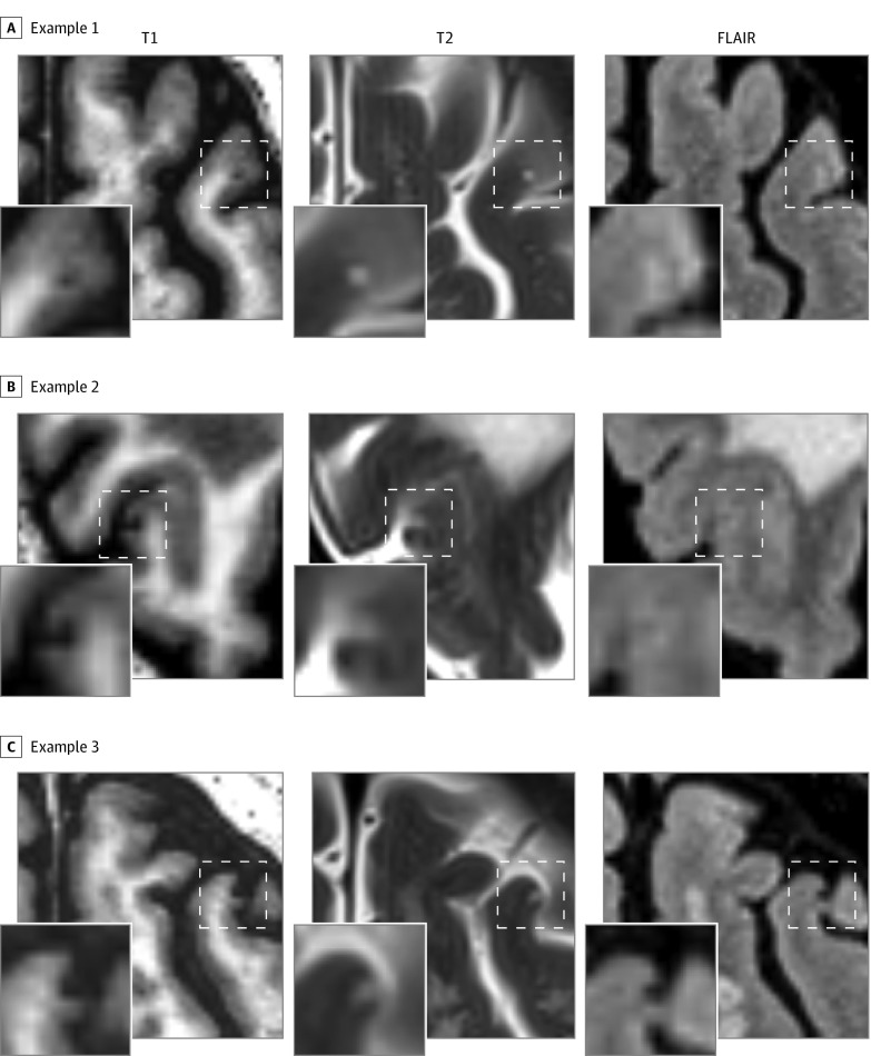
Figure 1. Examples of Chronic Cortical Microinfarcts on High-Resolution 3-T Imaging.
Chronic cortical microinfarcts were defined as hypointense lesions on T1-weighted imaging in combination with a hyperintense or isointense signal on T2 and a visible signal alteration on fluid-attenuated inversion recovery (FLAIR). A and B, The microinfarct is characterized on FLAIR by a hyperintense signal, whereas in panel C the signal is hypointense.

