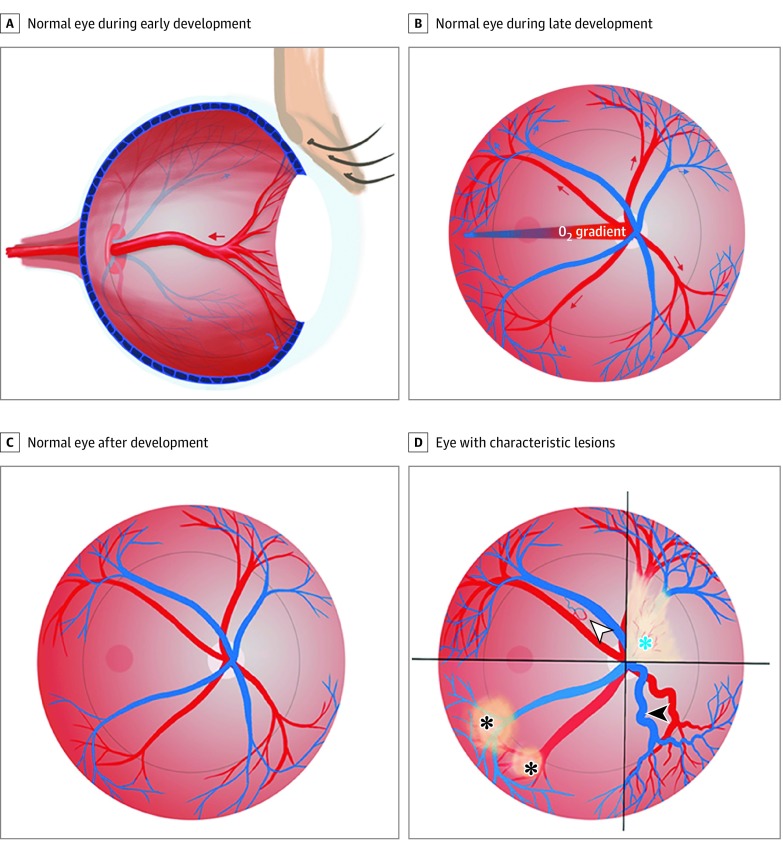
Figure 4. Schematic Illustrations.
A, The hyaloid artery supplying the lens is shown in a normal eye during development. As development continues, the hyaloid artery regresses (red arrow). B, A pattern of vessel growth is shown in a normal eye during development. As the arteries grow out from the optic disc and branch in the radial plane (red arrows), the increasing oxygen tension and decreased hypoxia signaling, mediated by hypoxia-inducible factor 2α (HIF-2α), causes the plexiform venous bed to regress to the periphery (blue arrows). C, When development is complete, the normal retinal vessel pattern is characterized by vessels that have emerged from the optic nerve head and spread, in a radial fashion, to meet the ora serrata in the periphery. D, The 4 characteristic lesions associated with a common HIF-2α gain-of-function-mediated disturbance of development are shown: fibrovascular membrane overlying the optic disc (blue asterisk), dilated and tortuous retinal vessels (white arrowhead), retinal pigment epithelium changes (black asterisks), and arteriovenous shunt (black arrowhead).

