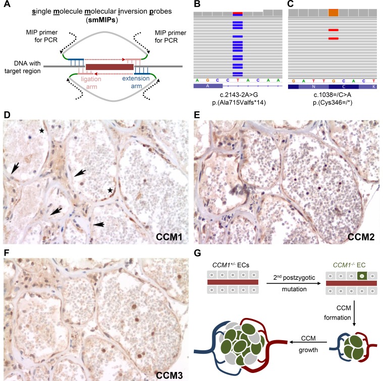
Figure 1.
Late postzygotic CCM1 nonsense variant and endothelial cell mosaicism within the cerebral cavernous malformation tissue from a proband with a de novo CCM1 germline mutation. (A) Genomic target regions were enriched and amplified using the smMIP technology. Hybridisation of smMIPs targeting each DNA strand, gap fill, ligation and binding sites for PCR primers are schematically depicted. Molecular tag sequences are marked in green. (B, C) Next generation sequencing analyses verified the heterozygous CCM1 germline mutation (B) and identified a postzygotic CCM1 nonsense variant (C). Endothelia of caverns presented with scattered immunopositivity and immunonegativity for CCM1 (D, arrows indicate negative and asterisks positive immunostaining), while CCM2 (E) and CCM3 (F) were uniformly present in serial sections. (G) Model of clonal evolution of an EC that acquired a second postzygotic mutation (CCM1−/−) and incorporation of ECs with an intact CCM allele (CCM1+/−) into the growing CCM. CCM, cerebral cavernous malformation; ECs, endothelial cells; smMIP, single-molecule molecular inversion probe.

