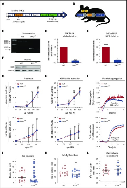
Figure 1.
Deletion of platelet IKK2 does not alter their function. (A) Graphical scheme of exons and their functional sites targeted by exon 3 and exon 6 and 7 deletion of IKK2. (B) Genetic scheme of IKK2ΔPlt mice. (C) Polymerase chain reaction (PCR) of WT megakaryocyte DNA of exon 3 and parts of adjacent introns generated an ∼2400–base pair (bp) band, whereas PCR of IKK2ΔPlt megakaryocyte DNA, with exon 3 deletion, generated an ∼730-bp band. (D) Quantitative PCR (qPCR) of an intron sequence between exon 2 and 3 of IKK2 that is excised during Cre recombination, normalized to a GAPDH intron sequence (n = 3). (E) qPCR of exon 3 of IKK2 messenger RNA, normalized to HPRT (n = 3). (F) Western blot of IKK2 and GAPDH (as loading control) of washed WT and IKK2ΔPlt platelets. P-selectin (G) and activated GPIIb/IIIa (H) median fluorescence intensity (MFI) of CD41+ events in diluted whole blood stimulated with PAR4-AP (upper panels) or CVX (lower panels) (n = 12). (I) Representative light-transmission aggregometry curves of washed WT and IKK2ΔPlt platelets, stimulated with 165 mU/mL thrombin (upper panel) or 18 µg/mL collagen (lower panel). (J) Tail tips (5 mm) of WT and IKK2ΔPlt mice were cut, and time until cessation of bleeding was recorded (n = 12-18). The horizontal line represents the median. (K) Mesenteric arteries of WT and IKK2ΔPlt mice were treated topically with 1.0 M FeCl3, and time until vessel occlusion was determined by intravital microscopy. Each symbol represents a mesenteric arteriole (n = 12-13). The horizontal line represents the median. (L) Recruitment of peritoneal macrophages after intraperitoneal injection of 4% thioglycollate. After 3 days, cells were harvested and analyzed by flow cytometry (n = 8-10). Dashed line represents basal count of untreated WT mice. **P ≤ .01, ****P ≤ .0001. ns, not significant.

