Fig. 1.
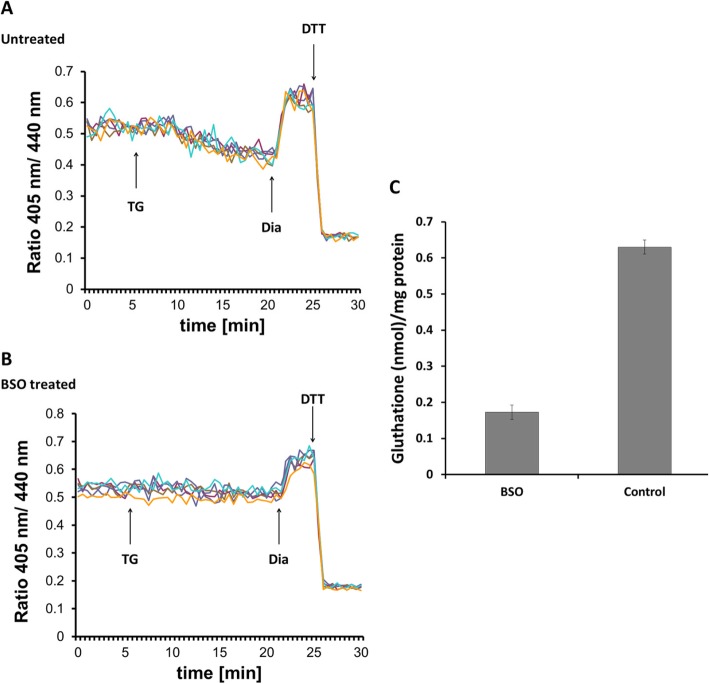
Ca2+ depletion-triggered ER reduction is sensitive to glutathione depletion by BSO. HEK293 cells were stably transfected with Grx1-roGFP1-iEER constructs and subjected to ratiometric laser scanning microscopy on a temperature-controlled stage with CO2 control. Fluorescence ratio changes were monitored over time. Each trace corresponds to the data recorded from one cell; traces were obtained from two independent experiments. One micromolar TG were applied to untreated (a) or BSO-treated (b) cells as indicated by the arrow. At the end of each experiment, 500 μM diamide (Dia) and 20 mM DTT were added to ensure the functionality of the probe. c Determination of total glutathione concentration by glutathione reductase assay as described in the “Materials and methods” section. One millimolar BSO treatment was performed overnight prior to experiment

