Fig. 2.
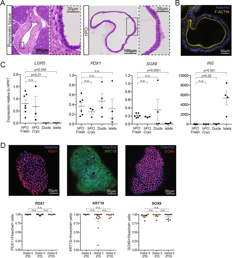
Human pancreatic organoids (hPOs) expanded long-term recapitulate pancreatic ductal epithelium in vitro. a Representative images of H&E staining of human pancreatic ductal tissue and hPOs. Note that hPOs (right) expanded in culture retain the single-cell morphology exhibited by the pancreatic ductal tissue in vivo (left) (n = 6 independent donors). b Representative immunofluorescence staining of F-Actin (yellow) demonstrates that hPOs maintain the epithelial cell polarity typical of ductal tissue (nuclei counterstained with Hoechst, blue) (n = 6 independent donors). c mRNA expression analysis of key genes involved in stem cell biology (LGR5), pancreatic fate (PDX1), ductal fate (SOX9) and β-cell function (INS) in hPOs derived from fresh tissue (hPO Fresh, n ≥ 6), cryopreserved tissue (hPO Cryo, n = 3), isolated primary ducts (n = 4) and isolated islets (n = 4). d Immunofluorescence staining (upper panel) and quantification of positive cells (lower panels) of nuclear PDX1 (red), cytoplasmic KRT19 (green) and nuclear SOX9 (red) protein in hPOs. Graphs represent number of positive cells for the corresponding marker (≥7 organoids counted per donor). Graphs show mean ± SEM

