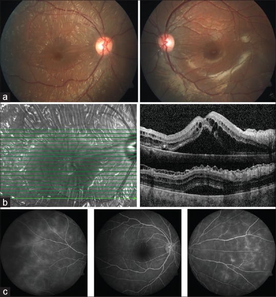
Figure 1.

(At Presentation) (a) Fundus photograph in OD shows glistening cellophane retinal reflexes with folds in posterior pole up to equator. OS has retinal sheen. (b) SD-OCT in OD shows abnormal internal limiting membrane, schitic spaces between ILM, and Nerve fiber Layer, intraretinal schisis with serous detachment at macula (c) Fluorescein angiography in OD reveals small vessel leak in late phases
