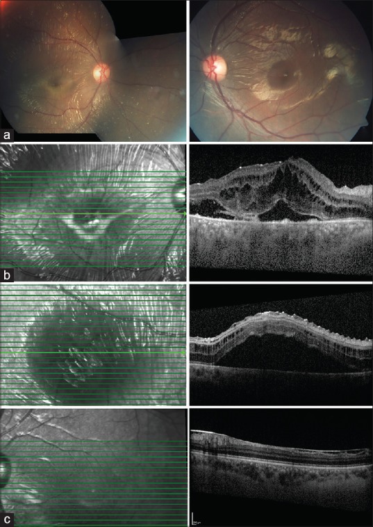
Figure 2.

(At 4 months follow up) (a) OD has retinal reflexes, folds with subretinal exudation (b) SD-OCT in OD has increased Intraretinal schisis with a new area of serous detachment temporal to macula (c) OS has epiretinal membrane in inferior macula
