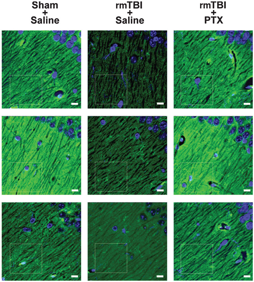
Fig. 6.
Paclitaxel treatment blocks rmTBI-induced loss of postsynaptic density-95 immunoreactivity in hippocampus. Representative PSD-95 (green) expression in CA1 stratum oriens hippocampi. Tissue was counterstained with DAPI (blue) to delineate prominent nuclei of the stratum pyramidale. In saline-treated mice exposed to rmTBI, hippocampal PSD-95 immunofluorescence was reduced as compared to sham or rmTBI paclitaxel mice. CA1 (just superior to the dentate lateral horn) is near the location of the Gallyas silver stain from Fig. 2C. Confocal micrographs are from 3 mice in each group. White boxes indicate region of fluorescence analysis. Bars = 10 μm.

