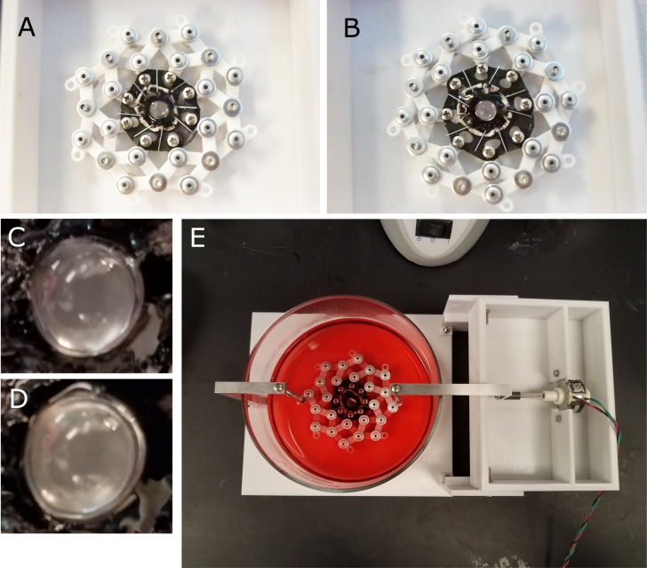
Figure 1.
The anterior portion of the eye was isolated, and the cornea and iris removed; leaving the lens, ciliary body, and a ring of sclera intact. Eight flaps were cut into the sclera and each flap was affixed to the silicone disk using staples. The silicone disk was mounted onto the stretching ring which, when expanded, would stretch the lens equally along eight axes. Disk mounted onto stretching ring in unstrained (A) and strained (B) configurations. A close up of the lens in the unstretched (C) and stretched conditions (D). The lens and accessory tissue mounted onto the motorized lens stretching device and submerged in culture media (E). The amplitude and frequency of the lens stretching regime can be customized and run for the entire duration of the tissue culture period.

