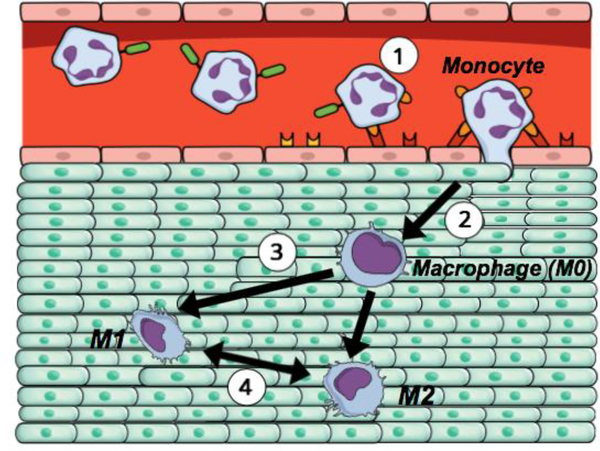
Figure 1.
The schematic representation of a monocyte extravasation into a tissue and macrophage polarization (1) A circulating monocyte from the bloodstream attaches to the endothelial layer and begins to infiltrate into the targeted tissue, (2) Upon extravasation, monocyte differentiates into macrophage (M0), which further exposes to the chemical, physical, and mechanical cues experienced within the targeted tissue, (3) Macrophage polarizes into classically activated (pro-inflammatory; M1) and alternatively activated (anti-inflammatory; M2) phenotype depending on the tissue milieu, (4) Macrophage differentiation is temporal. Macrophage may change its polarization state by time depending on the needs of the tissue.

