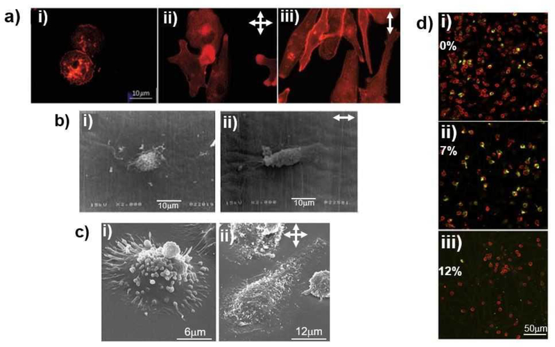
Figure 3.
Mechanical strain-dependent changes in macrophage morphology (A, B, and C) and phenotype (D). (A) Fluorescence images of U937 human macrophages upon without mechanical strain (i), biaxial (ii) and uniaxial (iii) mechanical strain exposure (98–100), (B) Scanning electron microscope (SEM) images of murine macrophages cultured under without mechanical strain (i) and uniaxial mechanical strain (ii) conditions (96), (C) Magnified SEM images of U937 human macrophages cultured under without mechanical strain (i) and biaxial mechanical strain (ii) conditions (96), (D) Fluorescence images of human macrophages and its phenotypic state (M1 stained in red and M2 stained in green) under various uniaxial mechanical strain magnitude; 0% (i) , 7% (ii), and 12% (iii) (101). For images at A,B, and C, the arrows indicate the direction of applied mechanical strain.

