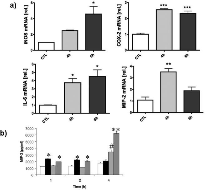
Figure 4.
The mechanical strain and its exposure time mediated mRNA level of proinflammatory genes in peritoneal macrophages (A) and MIP-2 protein secretion in fetal lung cells (B). (A) The changes in iNOS, COX-2, IL-1β, MIP-1α, and MIP-2 mRNA level with prolonged 20% mechanical strain exposure. * indicated p<0.05, ** indicated p<0.01, *** indicated p<0.001 versus unstretched controls. Error bars represented standard deviation with n=3. (49) (B) MIP-2 production of rat macrophages over time. White columns indicate control, Black columns indicated mechanical loading of 5% and 40 cycles/min, Light grey indicated LPS stimulation, and Dark grey columns indicate mechanical loading of 5% and 40 cycles/min and LPS stimulation. * indicated p<0.05 versus control, ** indicated p<0.05 versus all other groups, # indicated p<0.05 versus the rest of the groups (104)

