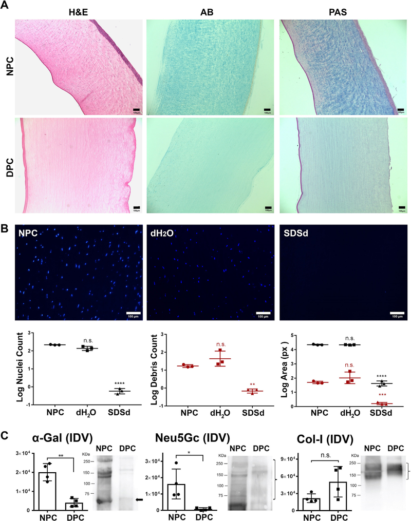
Fig. 1.
Assessment of decellularization efficiency. (A) Histological evaluation by staining with Hematoxylin and Eosin (H&E), Alcian Blue (AB) and Periodic Acid Schiff (PAS) on decellularized porcine cornea (DPC) compared with native porcine cornea (NPC). (B) Confirmation of decellularization by DAPI staining of nuclei and nuclear debris within NPC, corneas treated with distilled water (dH2O) and corneas treated with SDS in distilled water (SDSd). Graphs represent the nuclei (black color) and nuclear debris (red color) count and area covered by them. The results were reported as the mean ± S.D. Scale bars: 100 μm. (C) Specific detection of α-gal, Neu5Gc and collagen-I by Western Blot (WB) in NPC and DPC. The integrated density value (IDV) of each band was measured. Bar charts represent the calculated intensity of corresponding proteins using ImageJ densitometry. The results were reported as the mean ± S.D. from four independent corneas. Representative images of the WB are shown on the right (bands of interest are marked with arrow or bracket).

