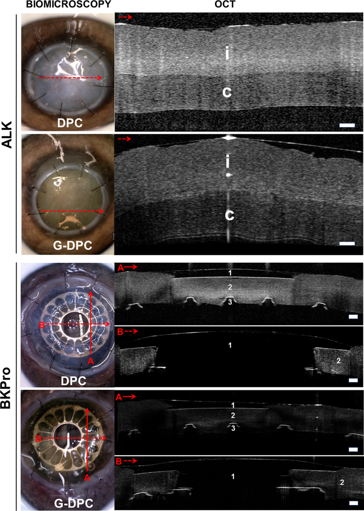
Fig. 8.

Biomicroscopy and OCT images of non-gamma (DPC) and gamma irradiated (G-DPC) decellularized porcine corneas transplanted ex vivo in pig eye as anterior lamellar grafts and as a carrier for BKPro. Red arrows on the slit lamp images represent the section and direction of the OCT. No gaps within implanted donor cornea (i) or host corneas (c) were observed in either model. 1, 2 and 3 represent BKPro front plate (made of PMMA), donor cornea and BKPro black plate (made of titanium), respectively. Scale bars: 200 μm.
