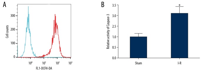
Figure 3.
The oxidative stress in the brain tissues of rats in the I-R group was apparent, and the activity of caspase-3 was significantly increased. (A) Flow cytometry was used to detect ROS content in the 2 groups of rats. (Green line is sham group and red line is I-R group) (B) The caspase-3 enzyme activity in the 2 groups of brain tissues was detected by spectrophotometry. * P<0.05 compared with the sham group. Each experiment was repeated 3 times. Compared with the sham group, the ROS content in brain tissue of the I-R group increased significantly (A). The results of spectrophotometry showed that compared with the sham group, the activity of caspase-3 in the brain tissues of I-R group increased significantly (B). I-R group – ischemia-reperfusion damage model; ROS – reactive oxygen species.

