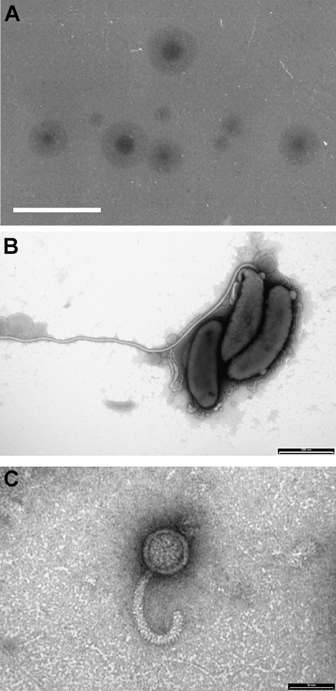
FIG 1.

Unique haloed plaque morphology from which the coisolated novel B. bacteriovorus angelus and bacteriophage halo were identified by electron microscopy. (A) Haloed plaques containing both B. bacteriovorus angelus and bacteriophage halo on lawns of E. coli in YPSC double-layer agar plates. Scale bar, 1 cm. (B) Electron microscopy of B. bacteriovorus angelus, stained with 0.5% URA (pH 4.0). Scale bar, 500 nm. (C) Electron microscopy of a 0.22-μm filtrate of a predatory culture, showing the presence of phage particles with curved tails resembling bacteriophage RTP. Phage were stained with 0.5% URA pH 4.0. Scale bar, 50 nm.
