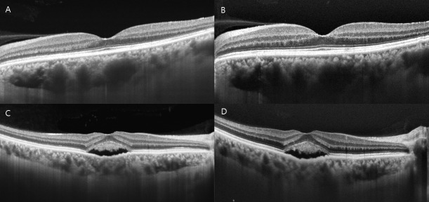
Fig 4. Representative optical coherence tomography (OCT) images showing the difference in the clarity of the choroid-scleral boundary between swept source (SS) and spectral domain (SD) OCT.
SS-OCT scan (A) and SD-OCT scan (B) of a patient with resolved central serous chorioretinopathy (CSC). The choroid-scleral boundary is observed more clearly in A than in B. SS-OCT scan (C) and SD-OCT scan (D) of a CSC patient with subretinal fluid (SRF). The choroid-scleral boundary of D is faint behind the SRF because of a shadow and hyperreflective layer within the choroid, whereas C shows a relatively clear choroid-scleral boundary.

