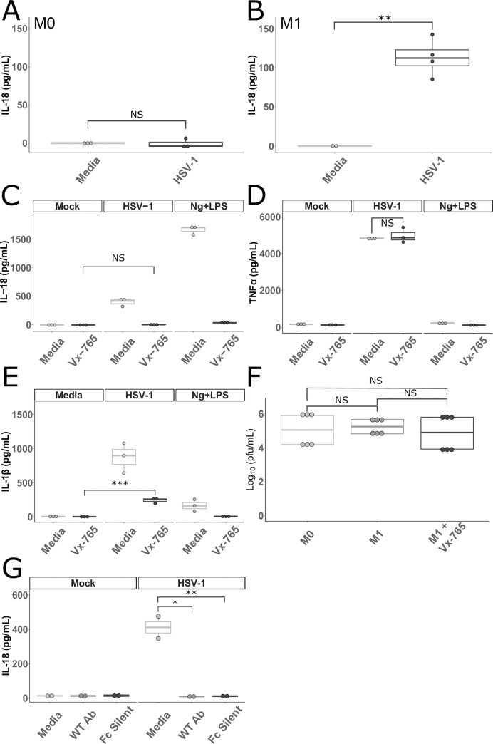
Fig 1. HSV-1 activates inflammasomes in primary human macrophages.
A and B. Primary human MDMs cultured without (M0 A) or with IFNγ (M1 B) were incubated with HSV-1 or media for 24 hours. Cell culture supernatants were collected and assayed for IL-18 (B represents the combination of two experiments). C, D, and E. Primary human MDMs stimulated with IFNγ were cultured in media alone or in media containing 100 μg/mL of VX-765 (Invivogen, San Diego, California) and then incubated with HSV-1, nigericin and LPS (Ng+LPS), or media for 24 hours as outlined in Materials and Methods. Cell culture supernatants were collected and assayed for (C) IL-18, (D) TNFα, and (E) IL-1β. F. MDMs cultured without IFNγ (M0), with IFNγ (M1), or with IFNγ and VX-765 (M1+Vx-765) were infected with HSV-1 for 1 hour followed by citrate wash to inactivate any extracellular virus. Supernatants were collected 24 hours later and plaque forming units (PFU) were determined via standard plaque assay on Vero cells. Data shown are combined from two independent experiments. G. MDMs stimulated with IFNγ (M1) were incubated with HSV-1 or media as well as a neutralizing antibody (WT Ab) and a neutralizing antibody unable to bind to Fc receptors (Fc Silent) for 24 hours. Cell culture supernatants were collected and assayed for IL-18. Differences between groups indicated by brackets were determined by a Student’s t-test. NS, *,**,*** indicate p-values >0.05, <0.05, <0.01, <0.001, respectively. The lower and upper borders of the boxplots represent the 1st quartile and 3rd quartile respectively. The median is represented by a horizontal line in the box. The lower and upper whiskers represent 1.5x the interquartile range (IQR) beyond the quartile lines. Each dot represents an individual sample.

