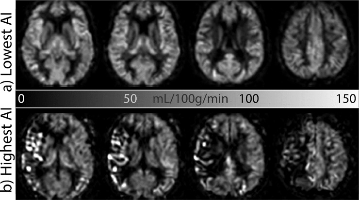
Fig 2. CBF images from the patients with the lowest (a) and highest (b) asymmetry index (AI) of the spatial coefficient of variation, sCoV.
Compare the overall symmetric and homogeneous appearance of the gray matter perfusion of (a) with the asymmetric, heterogeneous, vascular appearance of the apparent gray matter perfusion of (b). CBF = cerebral blood flow, sCoV = spatial coefficient of variation.

