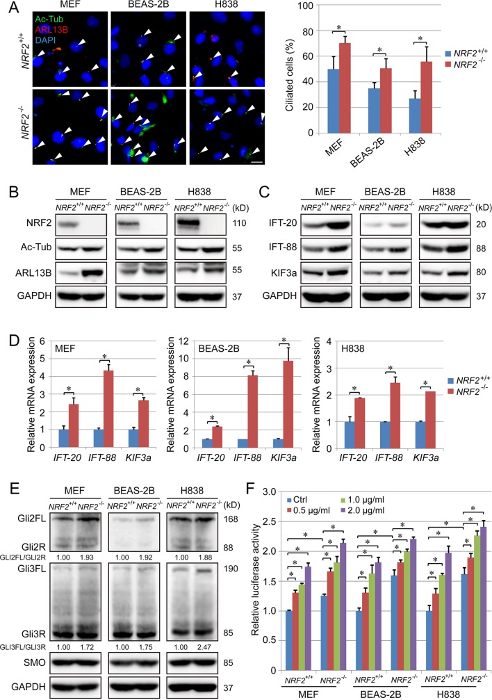
Fig 1. NRF2 deletion enhances ciliogenesis and Hh signaling.
(A) IF for Ac-Tub (green) and ARL13B (red) in NRF2+/+ and NRF2−/− MEFs, BEAS-2B, and H838 cell lines. Ciliated cells (%) represent the percentage of Ac-Tub/ARL13B-positive cells normalized to the total number of DAPI-positive cells in 6 random fields. (Scale bar = 10 μm.) (B) Immunoblot analysis of NRF2, Ac-Tub, and ARL13 protein levels in the indicated NRF2+/+ and NRF2−/− cell lines. (C) Immunoblot analysis of IFT-20, IFT-88 and KIF3a protein levels in the indicated NRF2+/+ and NRF2−/− cell lines. (D) qRT-PCR analysis of IFT-20, IFT-88 and KIF3a in the indicated NRF2+/+ and NRF2−/− cell lines. (E) Immunoblot analysis of GLI2, GLI3, and SMO expression in the indicated NRF2+/+ and NRF2−/− cell lines. (F) GLI luciferase assay in NRF2+/+ and NRF2−/− cell lines treated with 0, 0.5, 1, or 2 μg/ml Shh for 24 h. Relative quantification of immunoblot results is shown in S1A–S1C Fig. Results are expressed as mean ± SD. A t test was used to compare the various groups, and p < 0.05 was considered statistically significant. *p < 0.05 compared between the two groups. Ac-Tub, acetylated tubulin; ARL13B, ADP-ribosylation factor-like protein 13B; GAPDH, glyceradehyde-3-phosphate dehydrogenase; Hh, hedgehog; IF, immunofluorescence; IFT, intraflagellar transport; KIF3a, Kinesin Family Member 3A; MEF, mouse embryonic fibroblast; NRF2, nuclear factor-erythroid 2-like 2; qRT-PCR, Real-Time Quantitative Reverse Transcription PCR; Shh, Sonic hedgehog; SMO, smoothened.

