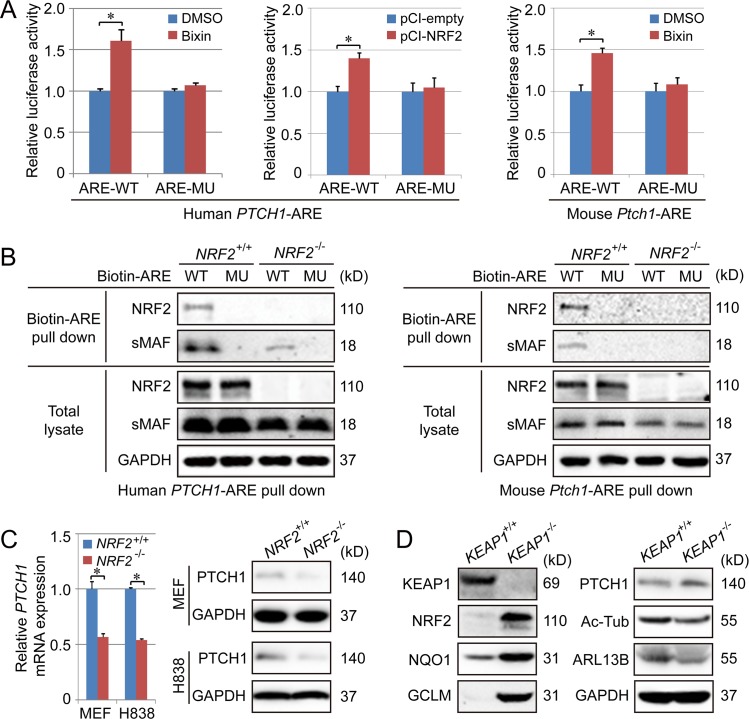
Fig 3. PTCH1 is an NRF2-target gene.
(A) H1299 and MEF cells were transfected with pGL4.22 vector containing the promoter of human PTCH1 or the promoter of mouse Ptch1 and further treated with bixin (40 μM) for 16 h, followed by a dual luciferase assay. (B) Biotinylated PTCH1-ARE-WT and PTCH1-ARE-MU were incubated with whole-cell lysates from H838 (for human PTCH1-ARE) or MEF cells (for mouse Ptch1-ARE), and ARE-bound proteins were pulled down by streptavidin beads and detected by immunoblot analysis with anti-NRF2 and anti-sMAF antibodies. (C) qPCR (left panel) and immunoblot (right panel) analysis of PTCH1 levels in the indicated NRF2+/+ and NRF2−/− cells. Relative quantification of immunoblot is shown in S6D Fig. (D) Immunoblot analysis of NRF2, NQO1, GCLM, Ac-Tub, ARL13B, and PTCH1 in KEAP1+/+ and KEAP1−/− H1299 cell lines. Relative quantification of immunoblot results is shown in S6E Fig. Results are expressed as mean ± SD. A t test was used to compare the various groups, and p < 0.05 was considered statistically significant. *p < 0.05 compared between the two groups. Ac-Tub, acetylated tubulin; ARE, antioxidant response element; ARL13B, ADP-ribosylation factor-like protein 13B; GAPDH, glyceraldehyde-3-phosphate dehydrogenase; GCLM, Glutamate-Cysteine Ligase Modifier Subunit; KEAP1, Kelch-like ECH-associated protein 1; MEF, mouse embryonic fibroblast; MU, mutation; NQO1, NAD(P)H Quinone Dehydrogenase 1; NRF2, nuclear factor-erythroid 2-like 2; PTCH1, Patched 1; sMAF, small MAF; WT, wild type.

