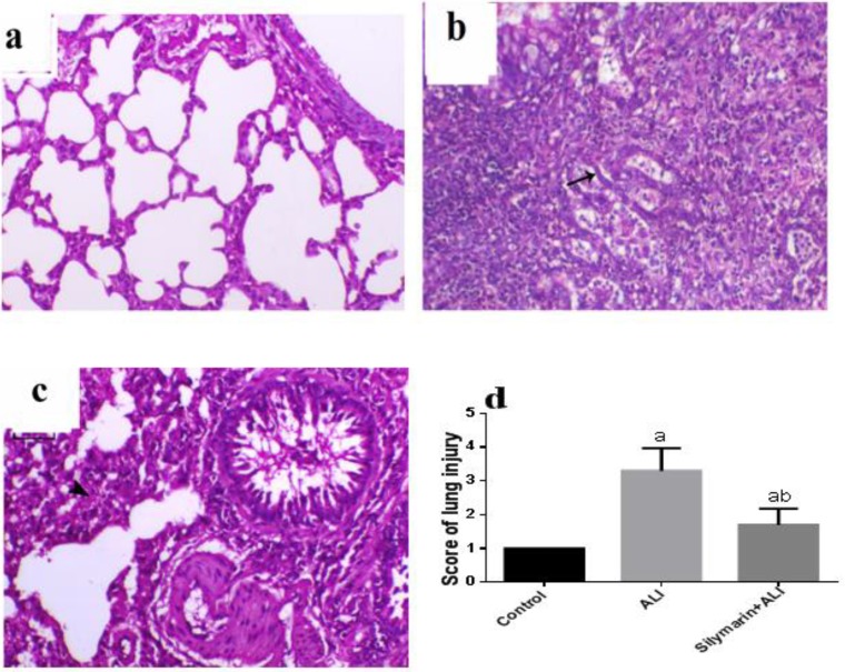
Figure 4.
Histopathology of lung tissue
Hematoxylin and eosin evaluation of rat lung (magnification x200): (a) Lung of normal animal showing normal alveolar and bronchial structures. (b) Lung of ALI animal showing epithelialization of the alveolar lining (arrow) associated with pneumonia and marked inflammatory cell infiltration either intra-alveolar or interstitial and heavily fibrous exudate deposition. (c) The lung of animal undergone lung injury and treated with silymarin showing moderately decreased interstitial thickening (arrowhead) and bronchial epithelium necrosis. (d) Semiquantitative scoring of lung injury. Values are representation of 10 values as means±SD. Results are considered significantly different when P<0.05. aSignificant difference compared with the control group, b significant difference compared with the ALI group. ALI: acute lung injury

