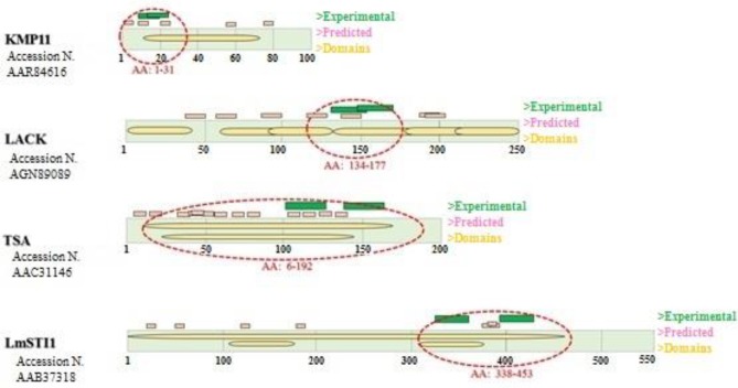
Figure 1.
Antigen structures with the number of protein sequence are shown in rectangles and domains within it. Experimental and predicted epitopes have been displayed in green and pink color, respectively above the rectangle. Selected Domains/oligopeptides in any antigen have been marked with red dash lines

