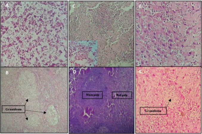
Figure 8.
Microscopic examination: Site of infection; diffuse macrophages with plenty of internalized amastigotes infiltrated with a few polymorphs and lymphocytes without or rare granuloma formation in the site of infection of control groups(A) and many granulomas with entrapped amastigotes in vaccinated groups (B). Spleen; visceral Leishmania infection in control groups’ mice altered the histological structure of the spleen, led to the loss of cell populations in the spleen, and disorganized architecture with indistinct border between red and white pulps (C) versus regular architecture in spleen of mice vaccinated with pleish-dom/pIL-12(D). Liver; immature granuloma formation with diffused amastigotes out of granuloma in liver sections of control groups (E) and the parasitized multinucleated granulomas seen in the liver sections of mice vaccinated with pleish-dom (F)

