. 2020 Jan 24;9:e52419. doi: 10.7554/eLife.52419

Figure 3. TH+ terminal patches are formed by a subset of cholinergic LOC intrinsic neurons that also express TH.

(A) A segment of the cochlea epithelium from a ChatiresCre; Ai3 mouse that labels cholinergic LOC efferents. Reporter protein EYFP is detected by an antibody against GFP. Cholinergic LOC fibers are found along the inner spiral bundle (ISB), under and around the IHCs. Cholinergic medial olivocochlear (MOCs) efferent fibers are found in the ISB and in the outer hair cell region. Co-labeling with TH immunostaining shows two patches of TH+ terminals (brackets) that overlap with a subset of the cholinergic terminals. TH also labels sympathetic fibers (SF). (B) Co-immunolabeling of TH and VAChT in the ISB of a wildtype mouse cochlea. A subset of VAChT-positive terminals are co-labeled with TH. (C) Two TH+ fibers with the characteristic morphology of LOC intrinsic neurons are shown in the cochlea of a 7-week-old old Th2a-CreER; Ai9 mouse with tamoxifen injection between 3–6 weeks of age (see Materials and methods). (D) Reconstruction of three sparsely labeled TH+ LOC neurons that show intrinsic neuron characteristics (axons in black; terminals in red). Two of the reconstructed fibers are shown in (C). The third one is from a different cochlea. See also Figure 3—figure supplements 1 and 2.

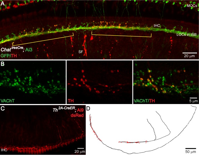
Figure 3.

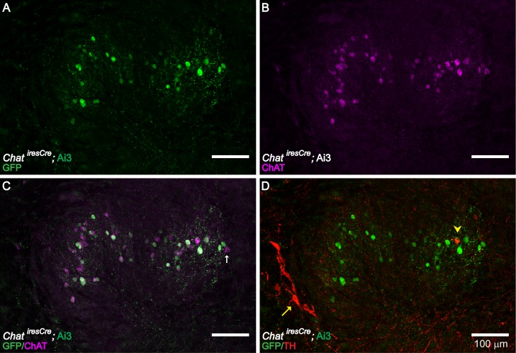
Figure 3—figure supplement 1. ChAT and TH Expression in the LSO Region of the Brainstem.

Figure 3—figure supplement 1.

ChAT immunolabeling confirms the cholinergic identity of LOC neurons genetically labeled in ChatiresCre; Ai3 mice (A–C) (n = 4 LSOs, three mice). Immunolabeling against TH (yellow arrowhead) labeled a small number of cholinergic LOC intrinsic neurons, as confirmed by co-labeling with ChAT antibody or ChatiresCre; Ai3 mice (D) (n = 41 LSOs, 23 mice). The previously described TH+, ChAT- shell neurons (Darrow et al., 2006b) are also observed (red arrow) (D) (n = 41 LSOs, 23 mice). (A–C) A brainstem slice from a P30 ChatiresCre; Ai3 mouse is immunolabeled against ChAT, demonstrating that LOC neurons labeled by the mouse line (a GFP antibody recognizes the reporter protein EYFP) are positive for ChAT. Note a small fraction of ChAT-immunopositive LOC neurons (magenta) were not labeled in ChatiresCre; Ai3 mouse (green) (arrow indicates one example), possibly due to different thresholds of reporter line and immunolabeling. (D) The same brainstem slice as in (A–C) is immunolabeled with TH, demonstrating the presence of a TH+/ChAT+ LOC intrinsic neuron (arrowhead) and a few of the previously described TH+/ChAT- LOC shell neurons (arrow) (Darrow et al., 2006a).
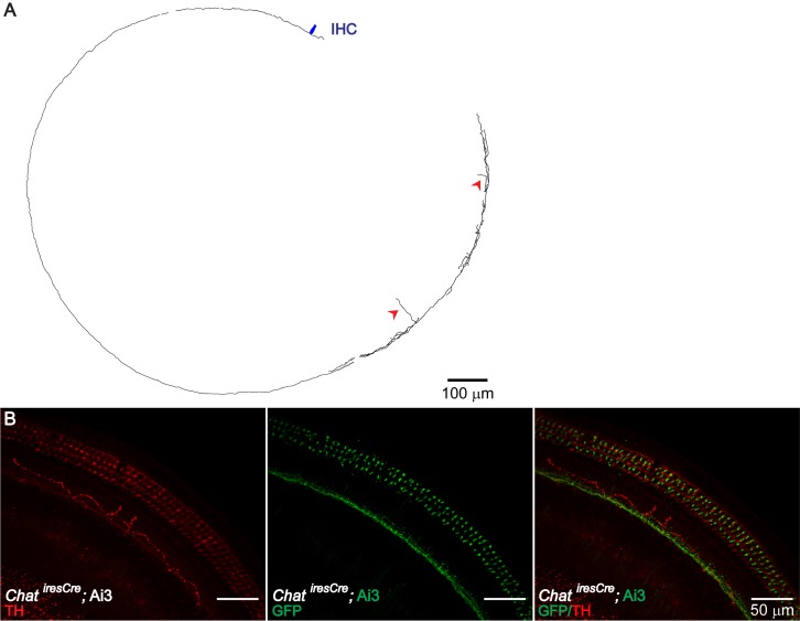
Figure 3—figure supplement 2. TH+ ChAT- LOC Fibers.

Figure 3—figure supplement 2.

Consistent with the previously reported TH+, ChAT- LOC shell neurons (Darrow et al., 2006b), LOC fibers with a typical shell-neuron morphology can be observed in sparsely labeled Th2A-CreER; Ai9 mouse cochleas (A) (n = 4, three mice). In addition, TH immunolabeling revealed another type of TH+ fiber, which travels around the IHC region in a meandering fashion (B) (n = 6 fibers, five mice). This type of fibers is not co-labeled by ChatiresCre; Ai3 mice (B) (n = 5 fibers, four mice). (A) Reconstruction of two LOC fibers labeled in a 7-week-old Th2A-CreER; Ai9 cochlea, both with a shell neuron-like morphology. The entry points into the organ of Corti for these two fibers are indicated with red arrowheads. One IHC is illustrated in navy blue for size comparison. (B) A cochlear segment of a P30 ChatiresCre; Ai3 mouse is immunolabeled with TH, demonstrating the peculiar meandering fiber path. This fiber is TH+ ChAT-; it does not overlap with the cholinergic fibers labeled by the mouse line.