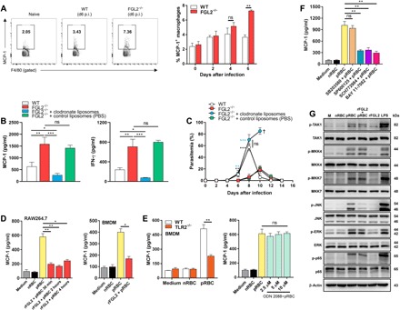
Fig. 4. sFGL2 inhibits parasite-stimulated macrophages from secreting MCP-1 through blocking JNK activation.

(A) Representative FACS (left) and statistical (right) analyses of the production of MCP-1 by macrophages from the spleens of both WT and FGL2−/− mice (n = 4) at the indicated time points. (B) The levels of MCP-1 (left) and IFN-γ (right) in the serum of the parasite-infected WT mice and FGL2−/− mice (n = 5) with or without macrophages depleted by clodronate liposomes at day 6 after infection. (C) The parasitemia of both parasite-infected WT mice and FGL2−/− mice (n = 5) with or without macrophage depletion by clodronate liposomes. (D) MCP-1 levels in the supernatants of pRBC lysate–stimulated RAW264.7 macrophages (left) and bone marrow–derived macrophages (BMDMs) (right) treated with or without rFGL2 for the indicated times as detected by ELISA. (E) MCP-1 levels released by WT and TLR2−/− BMDMs incubated with the pRBC lysate preparation as measured by ELISA (left), and the MCP-1 levels produced by the pRBC lysate–stimulated macrophages pretreated with or without the TLR9 antagonist ODN2088 as determined by ELISA (right). (F) MCP-1 levels in the supernatants of pRBC lysate–stimulated macrophages pretreated with or without nuclear factor κB (NF-κB) inhibitor BAY 11-7082, p38 inhibitor SB203580, c-Jun N-terminal kinase (JNK) inhibitor SP600125, and extracellular signal–regulated kinase 1/2 (ERK1/2) inhibitor SCH772984. (G) Western blots of the phosphorylation of TAK1, MKK4, MKK7, JNK, ERK1/2, and p65 in pRBC lysate–stimulated RAW264.7 cells pretreated with or without rFGL2. nRBC, normal red blood cell. Two to four independent experiments were performed. Data represent the means ± SD. *P < 0.05, **P < 0.01, ***P < 0.001. The symbol “†” indicates the time point when all mice died.
