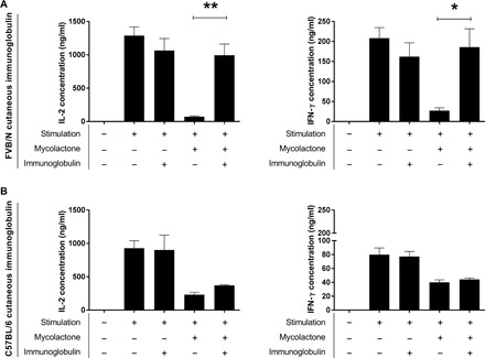
Fig. 3. Neutralization of mycolactone by cutaneous Ig from FVB/N mice infected with M. ulcerans.

CD4+ lymphocytes were stimulated with phorbol 12-myristate 13-acetate/ionomycin. Mycolactone was used at a concentration of 4 ng/ml. Igs purified from the skin of (A) FVB/N mice or (B) C57BL/6 mice infected with M. ulcerans were used at a ratio of 10 Ig molecules per molecule of mycolactone. Left: IL-2 quantification; right: IFN-γ quantification. The data shown correspond to one experiment performed in triplicate (means ± SD). *P < 0.05 and **P < 0.01 (Student’s t test).
