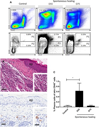
Fig. 4. Emergence of plasma cells (CD138+) in the skin during M. ulcerans infection in the FVB/N mouse model of spontaneous healing.

(A) Cells were analyzed by four-color flow cytometry with the specific staining of total lymphoid cells (CD45+), B cells (population P1 = CD45+, CD19+, B220+), plasmablasts (population P2 = CD45+, CD19+, B220int), and plasma cells (CD45+, CD19−, B220int, CD138+). The results shown here are from one representative experiment performed in duplicate. (B) Local inflammatory infiltration (inset) in the dermis of M. ulcerans infected mice at the ulcerative stage, typical of M. ulcerans infection. ep, epidermis. Immunostaining showing CD138+ plasma cells (activated B cells able to produce antibodies) in the dermis. (C) Flow cytometry analysis showing the emergence of plasma cells (CD45+, CD19−, B220int, CD138+) during the early steps of the spontaneous healing process (the control is uninfected skin). The histograms show the means ± SD. *P < 0.05 (Student’s t test). Scale bars, 160 μm (B top), 80 μm (B, top, inset), 100 μm (B bottom) and 20 μm (B bottom, inset).
