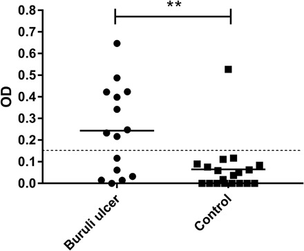
Fig. 5. Detection of cutaneous Igs binding to mycolactone in the lesions of patients diagnosed with Buruli ulcer.

Wells were coated with mycolactone, and antibodies binding to mycolactone in the plate were recognized by HRP-conjugated secondary antibodies. Biopsy specimens from patients with suspected and/or PCR-confirmed Buruli ulcer were provided by the CDTUB of Pobè (Benin). In PCR-confirmed Buruli ulcer patients, biopsies were performed on active lesions. It was not, therefore, possible to identify the patients subsequently displaying spontaneous healing. The detection limit was an absorbance of 0.150. **P < 0.05 (Mann Whitney U test). Buruli ulcer group, n = 15; control group, n = 19. OD, optical density.
