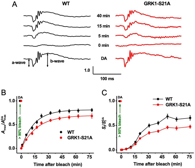
FIGURE 5.
Suppressed rod dark adaptation in GRK1-S21A mice. A, Representative scotopic ERG responses in the dark (dark-adapted [DA], bottom) and at four indicated time points after bleaching >90% of the rod pigment in wild-type (left) and GRK1-S21A (right) mice. For each time point, Amax values were normalized to their corresponding pre-bleach dark-adapted value (AmaxDA). B, Recovery of scotopic ERG maximal a-wave amplitudes (Amax; mean ± SEM) after bleaching >90% of rhodopsin in wild-type (n = 16) and GRK1-S21A (n = 15) mice. Bleaching was achieved by a 35-s illumination with bright 520-nm LED light at time 0. Averaged data points were fitted with single exponential functions yielding time constants of 14.0 ± 0.8 and 17.3 ± 0.9 minutes for wild-type and S21A-GRK1 mice, respectively. Final levels of response recovery by 75 minutes postbleach determined from exponential fits were 81% (wild-type) and 69% (GRK1-S21A). (C) Recovery of scotopic ERG a-wave flash sensitivity (Sf; mean ± SEM) after bleaching >90% of rod pigment in wild-type (n = 16) and GRK1-S21A (n = 15) mice. SfDA designates the sensitivity of dark-adapted rods. Animals and experimental conditions were the same as in B

