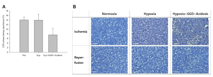
Fig. 3. Reperfusion damage under 1% serum condition.
Ischemia/reperfusion simulation was performed under 1% fetal bovine serum (FBS) conditions. The control group was cultured in DMEM containing 1% FBS for 24 h under normoxia. The hypoxia group was cultured for 24 h under hypoxia (1% oxygen). To simulate extreme ischemia, cells were cultured under hypoxia, glucose/glutamine deprivation (GGD), and lactic acidosis (pH 6.4). Cells were cultured for 24 h in these conditions. After the ischemic period, the medium was changed with DMEM containing 1% FBS and incubated under normoxia. Seventeen hours after the media change, lactate dehydrogenase (LDH) release was measured. Cell photographs were taken as soon as ischemia was over and as soon as the reperfusion was over. Data are expressed as means ± S.D. (n = 3). Scale bar: 200 µm.

