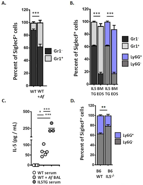
Figure 1. Mouse eosinophils express the Ly6G/Gr1 antigen.
A. Percent eosinophils (SiglecF+ cells) from bone marrow of wild-type (WT) BALB/c mice at baseline or sensitized and challenged with Aspergillus fumigatus (+Af) that are Gr1+ (light bar) vs. Gr1− (dark bar) as determined by flow cytometry with the mAb RB6–8C5; n = 3 – 5 per group, ***p < 0.005, Mann-Whitney u-test. B. Percent eosinophils (SiglecF+ cells) from bone marrow of BALB/c interleukin-5 transgenic (IL5TG) mice and from ex vivo cultures of from bone marrow-derived progenitors from BALB/c mice (BMEOS) that are Gr1+ (light bar) vs. Gr1− (dark bar). The cell surface antigen detected by RB6–8C5 was confirmed as Ly6G using mAbs 1A8 (anti-Ly6G, blue bar); n = 3 per group, ***p < 0.005, 1-way ANOVA; see also Suppl. Fig. 1. C. IL-5 (pg/mL) detected in serum and BAL of mice indicated in A. and B; *p < 0.05, ***p < 0.005, 1-way ANOVA. D. Percent eosinophils (SiglecF+ cells) from bone marrow of wild-type (WT) C57BL/6 (B6) and C57BL/6 IL-5 gene-deleted mice (B6 IL5−/−) that are Ly6G+ (blue bar) vs. Ly6G− (grey bar); n = 3 – 6 per group, **p < 0.01, Mann-Whitney u-test.

