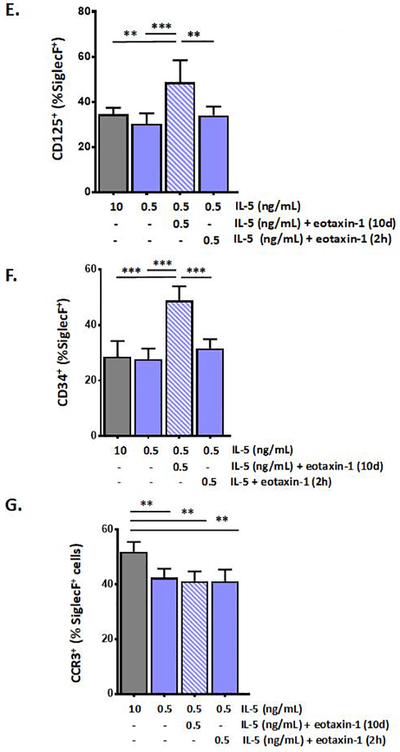
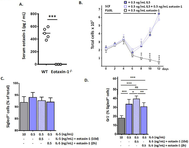
Figure 4. Eotaxin-1 limits expression of Ly6G/Gr1 on bone marrow-derived eosinophils (bmEos).

A. Serum eotaxin-1 in WT (wild-type) and eotaxin-1 gene-deleted mice; n = 3 – 5 mice, ***p < 0.005, Mann-Whitney u-test. B. Bone marrow-derived eosinophils cultures initiated under standard conditions as indicated; at day 4, these factors are replaced with 0.5 ng/mL IL-5 alone, 0.5 ng/mL IL-5 with 0.5 ng/mL eotaxin-1, or 0.5 ng/mL eotaxin-1 alone; total cells in culture on each day are as shown; n = 3 – 5 per point, ***p < 0.005. C. SiglecF+ cells (% of total) at day 12 of culture under growth conditions indicated; n = 5 – 8 per point; no significant differences detected between groups. D. Gr1− cells (% of SiglecF+) in bmEos cultures under growth conditions indicated; E. CD125+ (IL5-Rα+) cells (% of SiglecF+) in bmEos cultures as above. F. CD34+ cells (% of SiglecF+) in bmEos cultures grown as above. G. CCR3+ cells (% of SiglecF+) in bmEos cultures as above; for D. – G., n = 5 – 8 per point, *p < 0.05, **p < 0.01; ***p < 0.005, ns, not significant, 1-way ANOVA.

