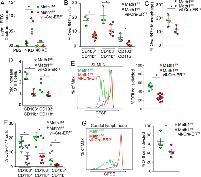
Figure 2: Goblet cells support antigen presenting cell acquisition of luminal antigen and CD4+ T cell responses to luminal antigen in the gut draining lymph nodes.
A) 4kD and 40kD FITC-dextran in serum after oral gavage in Math1f/fvil-Cre-ERT2 mice and Math1f/f littermate controls. B and C) Luminal Ova acquisition by SI LP-APCs assessed by flow-cytometric analysis two hours post oral gavage. D) Antigen presentation capacity of SI LP-APCs isolated from mice Math1f/f and Math1f/fvil-Cre-ERT2 mice given luminal Ova as assessed by expansion of Ova specific OTII T cells in ex vivo cultures. E) Histograms and quantification of in vivo proliferation of CFSE labeled OTII T cells in SI draining MLN of Math1f/f and Math1f/fvil-Cre-ERT2 mice 2 days after oral Ova gavage. F) Antigen acquisition by distal colon LP-APCs assessed by flow cytometry 2 hours following intra-colonic administration of fluorescent Ova. G) Histograms and quantification of in vivo proliferation of CFSE labeled OTII T cells in distal colon draining caudal LN of Math1f/f and Math1f/fvil-Cre-ERT2 mice 2 days after Ova enema. Data are representative of two or more replicates with ≥ 3 mice per group, each data point represents an individual mouse. Data is presented as mean +/− SEM, * = P < 0.05, ns = not significant.

