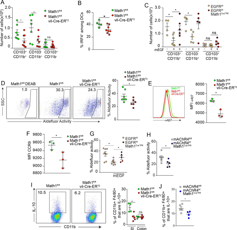
Figure 5: GAPs support the imprinting of LP-APCs.
Quantification of A) SI LP-APC subsets and B) IRF4+ SI LP-DCs in goblet cell deficient mice (Math1f/f vil-Cre-ERT2 mice) and littermate controls. C) Quantification of SI LP-APC subsets in mice lacking EGFR in goblet cells (EGFRf/fMath1Cre*PR mice) and littermate controls treated with mEGF. D) Flow cytometry dot plots and quantification of aldehyde dehydrogenase (ALDH) activity in LP CD11c+ MHCII+ SI APCs in goblet cell deficient mice (Math1f/f vil-Cre-ERT2 mice) and littermate controls. Expression of gut homing molecules E) α4β7 and F) CCR9 on OTII T cells following three days of in vitro culture with Ova and CD103+ DCs isolated from goblet cell deficient mice or littermate controls. Quantification of SI LP-APC with ALDH activity in G) mice lacking EGFR in goblet cells (EGFRf/fMath1Cre*PR mice) and littermate controls treated with mEGF and in SI LP-APCs from H) mice lacking mAChR4 in goblet cells (mAChR4f/f Math1Cre*PR mice) and littermate controls. I) Flow cytometry plots of SI LP macrophages and quantification of IL-10 expression by LP CD45+ CD11c+ MHCII+ F480+ cells from mice lacking goblet cells and their littermate controls. J) Quantification of IL-10 expression by SI LP macrophages from mice lacking mAChR4 in goblet cells and their littermate controls. * = P < 0.05, ns = not significant. Data is presented as the mean +/− SEM. Each data point represents and individual mouse, with the exception of E and F, where LP-APCs were pooled from three goblet cell deficient mice or three littermate controls.

