Figure 3.
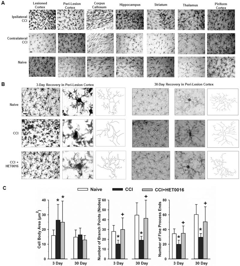
Morphologic changes of microglia after CCI. (A) At 3 days after CCI in rats, immunohistochemical staining for Iba-1 revealed a change in microglia morphology consisting of increased cell body size and less ramification of processes throughout the hemisphere ipsilateral to the injury relative to that in the corresponding contralateral regions and age-matched naïve brain. Scale bar = 20 μm. (B) Images of Iba-1-stained microglia in peri-lesion cortex at 3 and 30 days after CCI and in naïve rat cortex. Scale bar = 20 μm. Morphometric analysis of high-power images of microglial cell bodies and processes with Neurolucida software indicated that loss of process complexity persisted in vehicle-treated rats 30 days after CCI, whereas process complexity was better retained at 3 and 30 days in HET0016-treated rats. (C) Calculations of microglial cell body area, number of cell process branch points, and number of fine end processes (mean ± SD) in rat peri-lesion cortex at 3 and 30 days in naïve (3 days, n=8; 30 days, n=3), CCI + Vehicle (3 days, n=8; 30 days, n=17), and CCI + HET0016 (3 days, n=8; 30 days, n=10) groups. *P<0.05 versus naïve group; +P<0.05 versus CCI + Vehicle group (ANOVA and Bonferroni correction for multiple comparisons).

