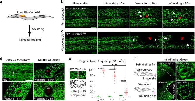
Fig. 1. Wounding triggers rapid and reversible mitochondrial fragmentation.
a Experimental design to investigate the mitochondrial response to epidermal wounding (laser or physical damage) in C. elegans. Mitochondria were labeled with matrix targeting sequence from cox 8 (mito) fused with XFP (including Pcol-19-mito::GFP(juEx4796), Phpy-7(y37a1b.5)-mito::GFP(yqIs157)70, Pcol-19-mito::mKate2(zjuSi47), or Pcol-19-mito::dendra2(juSi271)). b, c Laser wounding induces mitochondrial fragmentation in the epidermis. Representative confocal images of the epidermal mitochondrial network before and seconds after wounding by femtosecond laser (b) (see also Supplementary Movie 1, N = 5 independent experiments) and Micropoint UV laser (c) (see also Supplementary Movie 2, N = 7 independent experiments). Pcol-19-mito::GFP(juEx4796) was used to label mitochondria. We define mitochondrial fragmentation as a change from the interconnected tubular structure network to a rounded shape. d Mechanical needle wounding causes fragmentation of epidermal mitochondria, which return to normal morphology 24 hours after wounding except for a scar region at the center of the wound site. Representative confocal images of epidermal mitochondria before and after needle wounding. N = 3 independent experiments. Pcol-19-mito::GFP(juEx4796) was used to label mitochondria. Red asterisks in b–d indicate the wound site. White dashed squares indicate the zoom-in images for panel e. Scale bars b–d, 10 μm. e Quantitation of mitochondrial fragmentation frequency after needle wounding, measured in 100 μm2 regions of interest (white dash square in panel d) 10 μm adjacent to the wound site. Top panel shows enlarged images of mitochondria in unwounded (UW, n = 25) and wounded (W, n = 35) epidermis. Scale bars, 5 μm. Bars indicate mean ± SEM. ****P < 0.0001, Two-tailed unpaired t-test for wounded animals. Source data are provided as a Source Data file. f Wounding induce mitochondrial fragmentation in zebrafish tail fin. Left, experimental design for zebrafish tail fin wounding, 3 dpf larvae were first stained with mitoTracker Green for 2 h and then were wounded using needle. N = 3 independent experiments. Right, representative confocal image of mitochondria at the edge of zebrafish tail fin, Scale bar, 10 μm.

