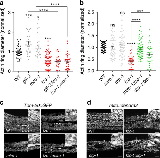
Fig. 5. Mitochondrial fragmentation acts downstream of Ca2+-MIRO-1 to accelerate wound closure.

a Loss of function in the fusion gene fzo-1 suppresses wound closure defects of gtl-2(n2618) and mcu-1(ju1154) mutants. Quantitation of actin ring diameter in WT and mutants 1 h after needle wounding (WT, n = 50; gtl-2, n = 27; mcu-1, n = 33; fzo-1, n = 64; gtl-2;fzo-1, n = 20; fzo-1;mcu-1, n = 33 animals). gtl-2 and mcu-1 mutants display delayed actin ring closure, which is suppressed in double mutants with fzo-1(tm1133). The WT actin ring diameter was normalized to 1 and mutants normalized to WT. Bars indicate mean ± SEM. *P = 0.0328, ***P = 0.0001, ****P < 0.0001, One-way ANOVA Dunnett’s test (single mutant versus WT). two-tailed unpaired t-test (versus mcu-1 or gtl-2). b Quantitation of actin ring diameter in the mitochondrial mutants (WT, n = 34; drp-1, n = 54; miro-1, n = 47; fzo-1, n = 44; miro-1;fzo-1, n = 82; fzo-1;drp-1 RNAi, n = 39 animals). drp-1 partially suppresses fzo-1 while miro-1 significantly suppresses the fzo-1 in enhanced wound closure. Bars indicate mean ± SEM. ns, P = 0.7753 (drp-1) or 0.9893 (miro-1), ***P = 0.0002, ****P < 0.0001, two-tailed unpaired t-test (versus fzo-1). One-way ANOVA Dunnett’s test (single mutant versus WT). Source data are provided as a Source Data file. c Representative confocal images of mitochondrial morphology in fzo-1 and miro-1;fzo-1 mutants. Pcol-19-Tomm-20::GFP(zjuSi48) transgenic animals label the mitochondria. fzo-1 mutants display fragmented and round shaped mitochondria. miro-1;fzo-1 double mutants have tubular mitochondria, similar to miro-1 single mutants. N = 3 independent experiments. d Representative confocal images of mitochondrial morphology in fzo-1 and drp;fzo-1 mutants. Mitochondria were labeled with mito::dendra2(juSi271), the mitochondrial morphology of drp-1;fzo-1 mimics drp-1, without fragmented mitochondria. Note, drp-1 suppress fzo-1 in mitochondrial morphology. Scale (c, d), 10 μm and 5 μm (zoom-in image).
