Figure 1.
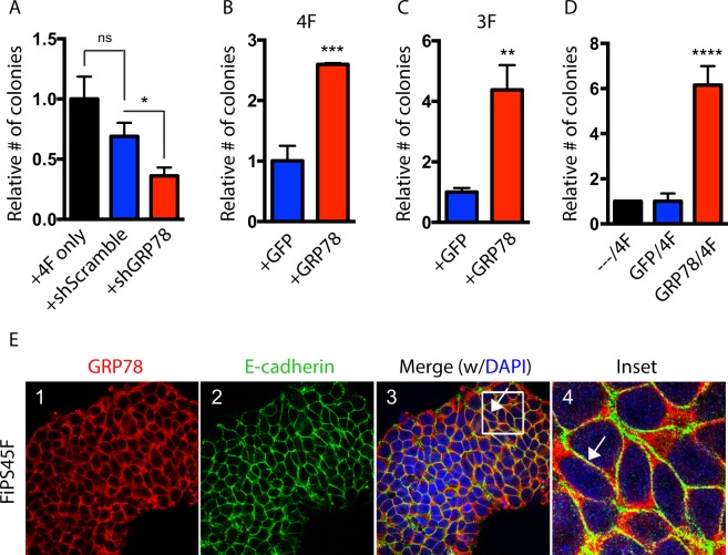
GRP78 is important for reprogramming and is expressed on the surface of iPSCs. (A) Human keratinocytes were retrovirally infected with OCT4, SOX2, KLF4 and c-MYC (OSKM) either alone (4F) or in the presence of shGRP78 or shScramble control. Resulting colonies (~20 days after infection) were stained for Nanog and colony numbers determined relative to the control. (B) Keratinocytes were retrovirally infected with OSKM (4F) or (C) with OSK (3F), in addition to a retrovirus expressing GRP78 or a GFP control. The number of Nanog positive colonies are shown relative to the control. (D) Keratinocytes were retrovirally infected with 4F following a 2-day prime with either a retrovirus expressing GRP78 or a GFP control. Nanog positive colony numbers are shown relative to the control. (E) iPSCs derived from fibroblasts (FiPS4F5) were examined by immunofluorescence for GRP78 (red), and E-cadherin (green, a cell surface marker), and both markers were found to colocalize together (Merge, Inset; arrows). 4,6-Diamidino-2-phenylindole (DAPI) staining shows nuclei. Results were quantified from triplicate samples, and are representative of at least three independent experiments. Error bars depict the standard error mean (SEM). A one-way ANOVA with a Tukey post hoc test (for multiple comparison tests) or an unpaired t-test (when comparing two samples) were used to determine statistical significance compared to controls, with p < 0.05 being considered statistically significant. *p < 0.05; **p < 0.01; ***p < 0.001; ****p < 0.0001; ns = not significant.

