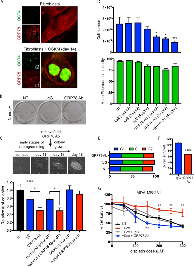
Figure 2.
Cell surface GRP78 plays a significant role in reprogramming and pluripotent stem cell and breast cancer cell function. (A) dFib-OCT4GFP fibroblast cells were plated on coverslips and left untreated (upper) or were infected with OSKM to initiate reprogramming (lower). 14 days post infection, cells were fixed and stained with OCT4 and GRP78. Day 14 shows the appearance of OCT4-positive cells (arrows, inset) at this timepoint, indicative of cells that have undergone reprogramming back to a pluripotent state. Note the change in GRP78 localization, primarily at the cell surface (insets). (B,C) Keratinocytes were infected with OSKM, plated, and cells were treated with media only, a GRP78 inhibiting antibody or IgG control throughout, or for the indicated timepoints. Following approximately 20 days after OSKM infection, resulting colonies were stained for Nanog (representative staining at this timepoint shown in B, used as the endpoint in the assay), and colony numbers determined relative to media only control. Note that cell surface expression of GRP78 (sGRP78) appears to be required for the reprogramming process (e.g. early anti-GRP78 timepoints affect colony numbers), rather than affecting colony number after colonies had already formed. Morphological pictures of cells throughout the reprogramming process (all at same magnification) are shown. (D) Pluripotent stem cells where GFP is driven by the OCT4 promoter were plated in triplicate and treated with media only, media containing GRP78 antibody, or respective IgG control, at the concentrations indicated. Media was replaced daily for four days. Cell counts were determined by trypan blue exclusion (upper panel). GFP expression (as a measure of OCT4 expression) was analyzed by flow cytometry (lower panel). (E,F) PSCs treated for 4 days with GRP78 antibody or IgG control (4μg/ml each) were analyzed for (E) cell cycle distribution determined by propidium iodide (PI) staining followed by flow cytometry analysis or (F) MTT-based survival analysis. (G) MDA-MB-231 breast cancer cells that overexpressed RFP-GRP78 under a doxycycline-inducible promoter, in the absence or presence of a GRP78 inhibiting antibody, were used in MTT assays to assess cisplatin-induced apoptosis. Note that the resistance to cisplatin-induced death that overexpression of GRP78 provides (red line) is dependent on cell surface expression of GRP78 (blue line). Results were quantified from triplicate samples, and are representative of at least three independent experiments. Multiple comparison statistical analysis of each cisplatin dose was calculated for each condition compared to relative NT sample dose. For all panels, error bars and p values are as described for Fig. 1.

