Figure 3.
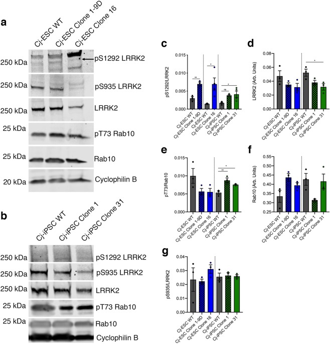
Marmoset LRRK2 kinase assay. (a) Representative Western Blot for pS1292 LRRK2 autophosphorylation, pS935 LRRK2, LRRK2, pT73 Rab10, Rab10, and cyclophilin B for Cj-ESC wild type (WT), Cj-ESC Clone 1–9D, and Cj-ESC Clone 16, and (b) Cj-iPSC WT, Cj-iPSC Clone 1, and Cj-iPSC Clone 31. (c) Relative quantification of pS1292/LRRK2 shows significantly increased pS1292 autophosphorylation in three G2019S clones compared to their respective wild type (WT) line. (d) LRRK2 protein expression levels (normalized to cyclophilin B) were consistent except for a significant decrease in Cj-iPSC Clone 31. (e) Relative quantification of pT73/Rab10 shows variability between Cj-ESC and Cj-iPSC lines but with significant increases in both Cj-iPSC G2019S clones. (f) Rab10 expression (normalized to cyclophilin B) was variable between all lines but without any significant difference. (g) There was no difference among lines for the constitutively phosphorylated pS935 LRRK2; n = 3–4 separately differentiated and collected samples per line. Note: artifact observed at the level of pS1292 detection was not quantified. (One-way ANOVA with Tukey’s multiple comparison was used to compare among Cj-ESC or Cj-iPSC lines. Student’s t-test was used for pS1292 in Cj-ESCs as the data for each G2019S clone was collected independently with respective WT controls; p < 0.05*; p < 0.01**).

